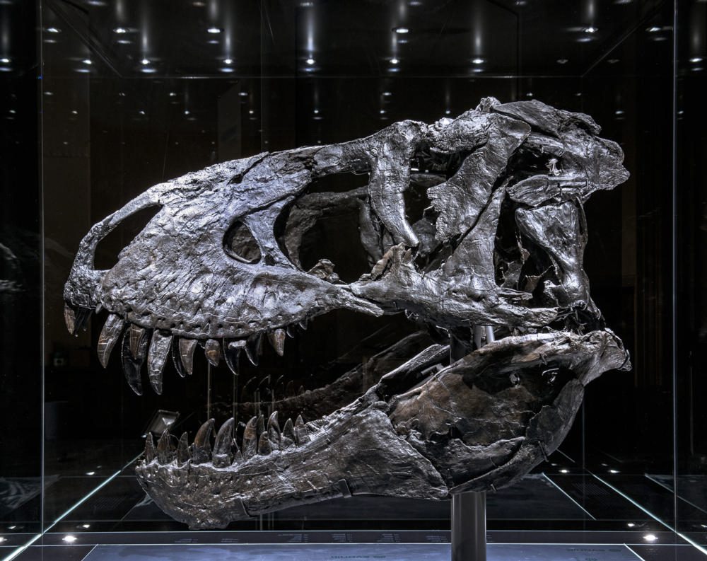
CT Uncovers Bone Disease in Tyrannosaurus Rex Jaw
The imaging method could have significant applications in paleontology, researchers said, as an alternative to fossil assessment methods that involve the destruction of samples.
The imaging method could have significant applications in paleontology, researchers said, as an alternative to fossil assessment methods that involve the destruction of samples.