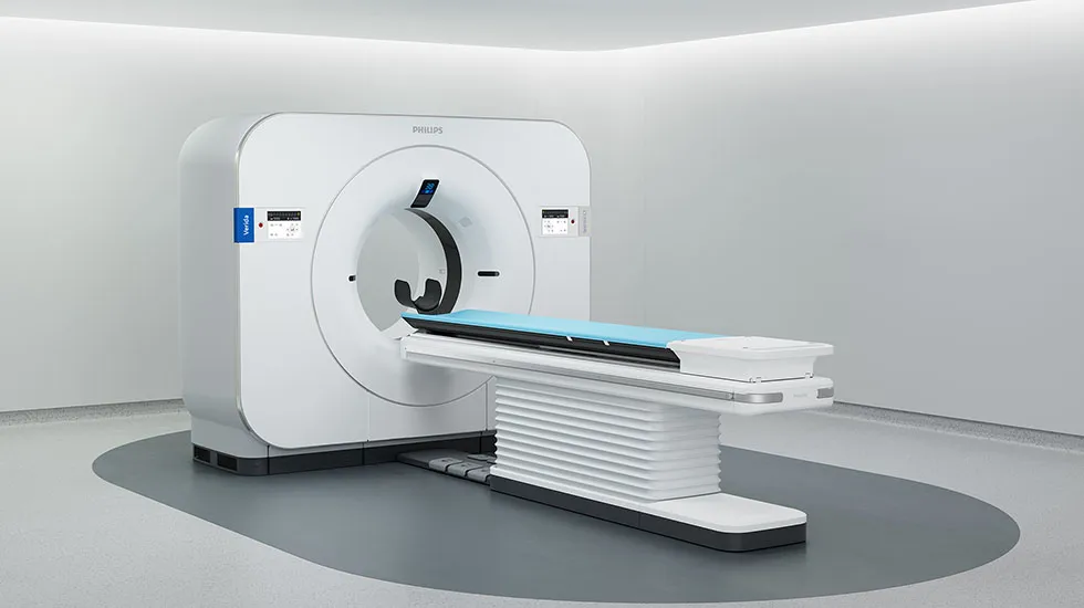
Royal Philips (NYSE: PHG, AEX: PHIA), a global leader in health technology, today announced it has received U.S. Food and Drug Administration 510(k) clearance for the Philips Spectral CT Verida system, bringing its next-generation, AI-powered spectral CT innovation to the United States.
AI-based reconstruction integrated into the imaging chain
Verida incorporates Spectral Precise Image, with a third-generation Nano-panel Precise dual-layer detector with intrinsic noise reduction, along with an AI-based deep learning reconstruction engine, designed to reduce noise and enhance image quality across CT applications [1]. Clinicians are able to customize optimal image de-noising according to their preferences.
“With FDA clearance for Verida, we are bringing the next evolution of spectral CT to more markets,” said Dan Xu, Business Leader of CT at Philips. “By combining always-on spectral imaging with AI-powered reconstruction, Verida enables clinicians to see more, first time right, supporting faster, more informed decisions and expanding the role of CT across clinical pathways.”
Always-on detector-based spectral CT for clinical versatility
Built on Philips’ industry-leading detector-based spectral CT technology, Verida enables always-on spectral imaging without requiring separate scans or workflow changes. It includes system and software enhancements designed to support clinical workflow, including improvements to the spectral result generation pipeline and updated computing infrastructure to support performance and usability. Verida reconstructs 145 images per second, so entire exams automatically appear in less than 30 seconds – 2× faster than previously, enabling up to 270 exams every day [2].
By capturing both high- and low-energy data in a single acquisition, clinicians can access conventional and spectral results simultaneously, supporting enhanced tissue characterization and material differentiation. This approach eliminates the need for pre-selection or repeat scans, enabling first-time-right imaging and deeper clinical insight across a broad range of applications [3].
Verida is a Computed Tomography X-ray system intended for diagnostic imaging in radiology, interventional radiology, and cardiology, and in oncology as part of treatment preparation and radiation therapy planning. It is indicated for head, whole body, cardiac, and vascular CT applications in patients of all ages [1]. The system is also intended to be used for low dose CT lung cancer screening when performed within established screening protocols [4]. Extended field-of-view images and respiratory correlated scanning (4DCT) are for treatment preparation and radiation therapy planning/simulation use only [3].
For more information about the Philips Spectral CT Verida system, visit the Philips website.