October 9, 2020
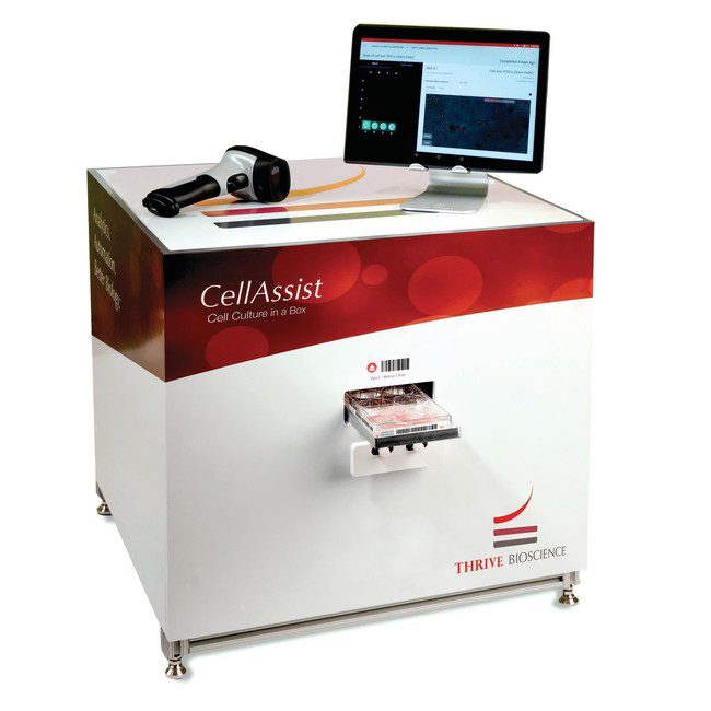
CellAssist which enables cell culture researchers to image, analyze, and document all cells, plates, reagents, and workflow details in a centralized database has been launched. The news was announced today. The CellAssist was developed by Thrive Bioscience, an innovator in cell culture systems, in collaboration with several highly-regarded academic institutions.
The CellAssist raises the bar for adherent cell culture by imaging and analyzing all the cells in a plate, not just cells chosen during typical manual inspection. Minutes after inserting a standard multi-well plate, the CellAssist captures, analyzes and displays hundreds of high-resolution images of cells at multiple magnifications, using phase contrast or bright-field. The visualization and analysis software allow researchers to comprehensively review images and metrics of the entire history of a project from one’s office or laboratory.
“The CellAssist solves cell culture problems widely experienced by researchers and laboratory managers, including inconsistent and partial imaging of plates of cells, a lack of objective metrics, and incomplete documentation,” said Thomas Forest Farb-Horch, Thrive Bioscience CEO and Co-Founder. “The CellAssist enables reproducible cell culture by capturing comparable images over time with barcoding and time-stamped documentation.”
“In cell biology, many of the important questions are raised after experiments are completed,” explained Steven Sheridan, Ph.D., Director, Platform for Cellular Modeling of Neuropsychiatric Disease, Center for Genomic Medicine, Massachusetts General Hospital. “With the CellAssist, we capture images and data, systematically and without the limitations that would later restrict our inquiries and insights. The database of images with related documentation becomes a valuable asset that helps us understand, analyze, and archive cellular metrics, unlike what we have previously achieved.”
Thrive Biosciences notes the CellAssist is available for shipment internationally.

