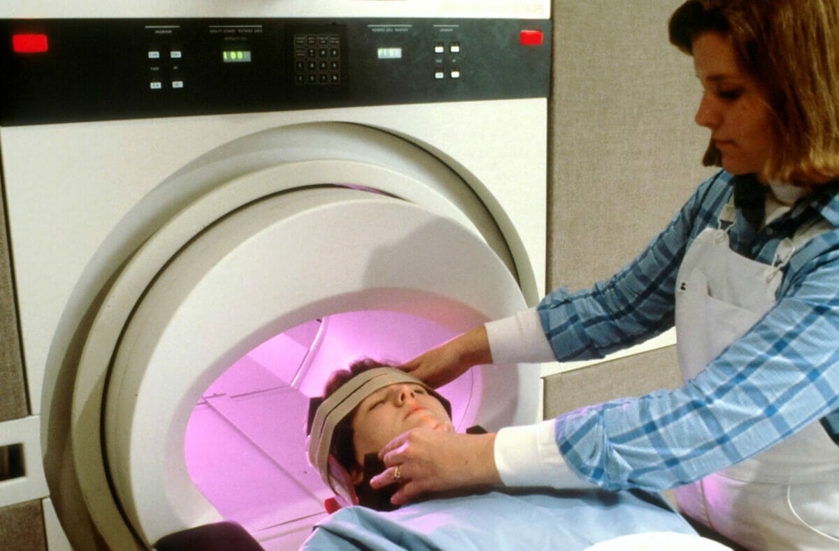
Medical imaging is a broad term that encompasses a variety of techniques used to create images of the internal organs and other structures within the body. These images can diagnose disease, plan surgery and treatment, or monitor patient progress after treatment. In many cases, medical imaging equipment works by using radiation to generate an image on film or digitally.
As healthcare professionals work with these advanced technologies, they often face challenges in understanding the intricacies and practical applications of various imaging techniques. To address these complexities, it is vital for professionals to collaborate and seek help with medical assignments, such as interpreting images or operating equipment. By fostering a collaborative environment, healthcare professionals can enhance their understanding of medical imaging technologies and ultimately deliver better patient care.
The earliest forms of imaging were X-rays and fluoroscopy (using visible light). Wilhelm Röntgen developed the first X-ray in 1895, but it wasn’t until about 50 years later that doctors could take images inside the human body without cutting them open with surgery. This led to many discoveries about how different diseases affect us internally—knowledge that previously made diagnosis difficult or impossible without invasive surgery. Since then, modern medical imaging equipment has become increasingly sophisticated for widespread use today, with certain manufacturers even using their expertise in aerospace composite manufacturing to provide the highest quality equipment available.
This led to many discoveries about how different diseases affect us internally—knowledge that previously made diagnosis difficult or impossible without invasive surgery. Since then, modern medical imaging equipment has become increasingly sophisticated for widespread use today.
Cost of Medical Imaging Devices
Here are some of the costs you are likely to encounter when purchasing new medical imaging equipment:
The cost of purchasing new equipment is high. New medical imaging devices can cost anywhere between $50,000 and $1 million. But if you buy used or refurbished equipment, you might save up to 70% instead of buying new. For example, an MRI scanner may cost approximately $2 million for a brand-new unit (and that price doesn’t include installation). However, by shopping for used items and refurbished parts instead of buying new ones directly from manufacturers (which often come with “bundled” services), healthcare providers can save up to 30% in cash outlay alone.
The cost of maintaining your machine is also high—it’s not cheap! Even if a device works perfectly well on its first day out of the factory, it still needs regular maintenance throughout its lifetime—something that most hospitals simply cannot afford in today’s economy; so again: used/refurbished machines make good sense here since they’re already broken-in before being sold.
Hence, instead of buying a new device, it is best to lease used imaging equipment to avoid upfront expenses and maintenance costs. Moreover, leasing also provides access to multiple tools within budget.
MRI Scanner
An MRI scanner uses magnetic fields and radio waves to create images of your body’s structures. It is a non-invasive, painless test that you can have done in about an hour. The most common uses for an MRI are:
- Diagnosing conditions such as brain tumors, stroke, and multiple sclerosis
- Assessing spine injuries or fractures (including stress fractures)
- Detecting tumors or masses in muscles, tendons, and ligaments throughout the body
CT Scanner (CAT)
A CT scan or CAT scan is a medical imaging technique that produces cross-sectional body images. These scans examine the bone and soft tissue, blood vessels, and organs.
CT scans use X-rays to create a picture of the inside of your body. The equipment that performs this test is a CT scanner (also known as an “X-ray machine”), which looks similar to an X-ray machine you may have seen at your dentist’s office or hospital.
A CT scanner uses a narrow beam of high-energy X-rays that rotate around your body, while an electronic sensor collects thousands of two-dimensional (2D) images from different angles. A computer then combines these 2D images into multiplanar reformats using software built into the machine itself or offloaded onto another computer via USB cable.
X-Ray
X-rays are generated by an X-ray tube, which is a device that uses electricity to produce beams of radiation. These beams pass through the body as they travel from one side to another. When they pass through soft tissues on their way, they are absorbed. However, when they pass through bone and other dense matter, the rays are reflected toward a detector in the machine that records them as an image.
X-rays can be used to look at bones, teeth, and lungs because these parts of the body contain little water and thus absorb less of the x-ray beam than soft tissues do. They are also beneficial for looking at the chest because it includes air (which absorbs quite a bit of x-ray energy) and bone (which does not absorb any x-ray energy). This makes it easy for radiologists to see what’s going on inside someone’s chest cavity without having any trouble getting enough contrast between different structures within that area of anatomy.
Ultrasound
Ultrasound uses sound waves to create an image. It is used to observe internal organs as they function, and it can guide a needle during a biopsy. It also assesses the flow of blood within vessels, the condition of muscles and tendons, and even the development of the fetus during pregnancy. Ultrasound is also used for other various purposes, including monitoring the growth and development of the fetus. One important ultrasound examination during pregnancy is the pregnancy dating scan, which helps determine the gestational age and estimated due date of the baby. Additionally, ultrasound is utilized in assessing the placenta’s position and function, as well as the amniotic fluid levels surrounding the baby. These ultrasound examinations provide vital information to healthcare providers, aiding in the comprehensive care and management of both the expectant mother and the developing fetus.
For ultrasound to work, your body must be penetrated with solid pulses of sound converted into electrical signals by tissue in your body (usually muscle). An electronic device then turns those electrical signals into images on a screen or monitor.
Ultrasound machines may have different capabilities depending on their purpose, but many have similar components: an electronic transducer that sends out high-frequency sound waves through water-filled gel; an amplifier that increases these signals so they can be detected by equipment; speakers that deliver them; computers for processing data and displaying images; monitors for viewing images (in color or black-and-white); keyboard entry devices for controlling what appears on the screen; printers for producing hard copies from digital files (elements from which medical reports are written); video recorders capable of storing data digitally so it can be transferred later via modem or USB cable connection.
Mammography Machine
Mammography is a diagnostic imaging technique that uses low-dose x-rays to produce images of the breast. The goal of mammography is to detect breast cancer in its earliest stages when it can be successfully treated. Mammograms are typically recommended for women over age 50, although younger women may need them if they have a family history of breast cancer or other risk factors.
Mammography machines are used to produce these images of the human breast, which are then analyzed by radiologists specializing in interpreting medical imaging results. While mammograms can be used for screening purposes—to check whether a woman has any abnormalities in her breasts—they’re often ordered as part of an evaluation after a suspicious nodule or lump has been found on an exam or self-exam.
Without imaging, doctors can’t see what’s happening inside the body. Hence, it has become one of the essential parts of modern medicine. Imaging helps doctors understand what may be going wrong and how to treat it.
