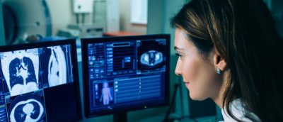
You, as a kid, must have daydreamed about the X-ray vision at least once in your lifetime. After all, seeing things that you can’t view normally sounds like a supernatural power in itself. In the current times, seeing something beyond a normal vision has a lot to do with practical applications and conducting treatments. All in all, it’s more than your superhero musings.
It’s all possible with the diagnostic imaging techniques, which demonstrate the finest work of radiologic medical treatments. It uses some uncommon approaches to save lives – all without a cape for sure!
What is diagnostic imaging?
It is a non-invasive diagnostic treatment. It means the practitioners can look through your body without conducting any surgery. The medical treatment involves different examination types to evaluate the inner organ functioning and movement. Generally, it is done to confirm the presence of diseases or internal injury. It can be seen as a crucial step in taking the medical proceedings further.
Radiologic specialists work closely with the patient to conduct tests appropriately. Here, slight negligence in tests can alter the results. Here are some of the aspects closely examined in the medical imaging approach – patient’s positioning, equipment protocol, test techniques, radiation protection, radiation safety, basic care of the patient, and much more.
Here are some common diagnostic imaging tests involved in this process –
#1 – X-rays – It is one of the most common types of medical examination performed to get maximum insights into the body. Some reasons where X-rays are highly recommended include – detecting the cause of pain, determining the injury, inspecting the disease progression, and much more. X-ray machines target a particular body part using radiation sensation to get visible insights.
#2 – MRI – It’s a preferred choice for the cross-sectional imaging process. MRI stands for magnetic resonance imaging. It is somewhat similar to a CAT scan. This examination type works the best for imaging soft tissues like – tendons and organs. Unlike the CAT scan, MRI does not use ionizing radiation but uses radio waves along with magnetic fields. It’s common for the patient to feel claustrophobic during the MRI process. As the examination is a bit noisy, the practitioner may fit earmuffs or earplugs to make you feel comfortable.
#3 – Ultrasound – Also referred to as sonography, ultrasound is ideal for capturing images within the body. This process uses high-frequency sound waves to give you a clearer picture of your body. The most common use of ultrasound is to detect health issues with soft tissues like vessels and organs. As it does not include radiation, it works as an ideal examination process for expecting mothers. The patients are advised to drink plenty of water before undergoing an ultrasound. This helps to obtain better pictures than usual.
The last word –

Most of the severe health issues begin with a thorough diagnostic imaging test process. This is significant to execute the treatment righteously. For the best results, it is advisable to contribute towards diagnostic treatment appropriately. Most of these examination types are pain-free and quick to give you test reports.
