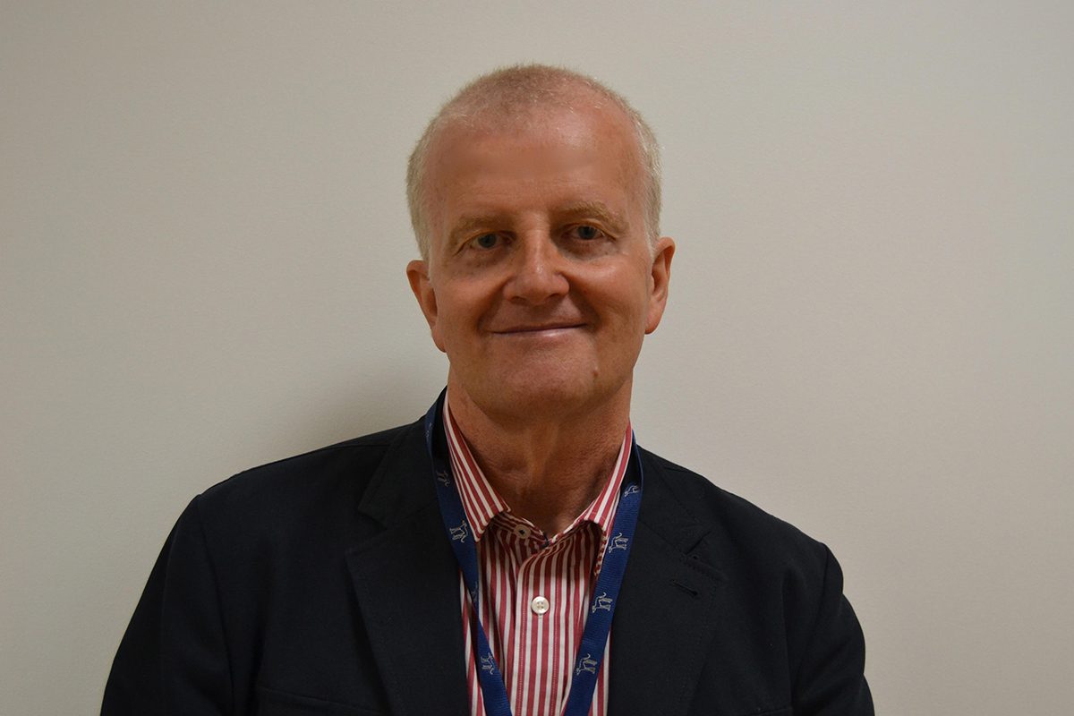
By: Professor Stefano Seri MD, FRCP, a Professor of Clinical Neurophysiology who regularly uses MEGIN technology
Despite its success in pre-surgical evaluation in localising brain activity, as well as being approved for the pre-surgical evaluation of medically refractory epilepsy, Magnetoencephalography (MEG) is often overlooked by physicians during the referral stage. Although studies and real-world patient outcomes continue to demonstrate MEG’s efficacy, clinicians still underrate its role in delivering valuable non-redundant information to help tailoring pre-surgical evaluation.
In this article, Professor Stefano Seri, Consultant Clinical neurophysiologist of the Epilepsy Surgery programme at Birmingham Children’s Hospital provides a contemporary look at how the imaging modality is optimizing diagnosis and changing paediatric and adult post-surgery outcomes for the better. Offering current thinking on the diagnostic modality’s efficacy, Professor Seri highlights the benefits of MEG within the approved indication, exploring how effective it is in supporting diagnosis, and surgical decisions pre-surgery.
Although MEG technology has been available to support the diagnosis and pre-surgical evaluation of epilepsy patients for more than 30 years, its uptake by clinicians has developed at a relatively slow pace. Results from a recent survey of European centres as well as a similar study conducted in the U.S., confirmed the inconsistent uptake (De Tiege et al., 2017). However, the speed of adoption may be accelerating as the weight of published evidence demonstrates the modality’s utility in the pre-surgical assessment of patients with drug-resistant epilepsy (Foley et al., 2019; Hall et al., 2018).
Time to reassess the cost/benefit ratio of MEG
Healthcare’s reluctance to adopt MEG more broadly into presurgical evaluation protocols may largely be attributed to outdated assumptions relative to the technology itself and its therapeutic value on patient outcomes including:
- The perception among clinicians that MEG data processing requires an army of in-house or outsourced physicists and engineers, limiting the “ownership” of the test by the clinical team at the point of care.
- MEG is more expensive and no more effective than other modalities capable of detecting and localising brain activity.
- The lack of a robust and internationally accepted analysis pipeline and standards that could potentially transform MEG into a widely available, economically efficient clinical tool.
- Absence of dedicated training and certification of EEG technologists and clinical neurophysiologists.
- Patchy, complicated healthcare reimbursement mechanisms that make financial planning challenging for healthcare providers.
By those that have a rudimentary understanding of MEG, it is still perceived as an interesting technology, with unclear benefits and delivering more return on investment in research rather than clinical application.
In 2011, a Canadian study looked at health technology assessment of MEG in presurgical assessment of children and adolescents with drug-resistant epilepsy. And concluded that the use of MEG prior to the initial multidisciplinary seizure conference resulted in a shorter time-to-surgical-candidacy decision compared to introducing MEG later, emphasizing the importance of early referral.
Although compelling scientific evidence of MEG’s efficacy is widely available, current guidelines reveal that it is not yet formally recommended.
MEG v. EEG: Contending for the gold standard in epileptic pre-surgical evaluation
When weighing the relative diagnostic utility and efficacy of MEG versus an electroencephalogram (EEG), certain practical aspects of the procedure highlight critical distinctions between the modalities and help elevate the clinical and therapeutic value of MEG scans above the alternatives.
Although high–density EEG recordings (128-256 channels) have a lower cost in terms of initial equipment investment, cost per test is not significantly different. For example, electrode caps and consumables used to record high-density EEG are expensive, their performance tends to degrade rapidly with use and the devices require specific attention in terms of infection control. The most expensive “consumable” of MEG systems is liquid helium which is available in limited supply. However, with advanced recovery systems available in many of today’s state-of-the-art patient-accessible systems, MEG has significantly reduced its operating costs.
Applying large number of electrodes on a patient is not without its challenges, particularly among those with learning or behavioural difficulties. Valuable MEG brain activity can be easily recorded in patients that categorically refuse to sit for a standard EEG. One of the advantages of MEG is that it requires less a-priori assumptions in modelling how brain currents flow inside the brain. Leading MEG system suppliers have steadily improved scanner designs, engineering in features that simplify the procedure for both technician and patient.
Benefits of MEG in comparison to fMRI
Functional magnetic resonance imaging or functional MRI (fMRI) measures brain activity by detecting changes associated with blood flow. The modality relies on the fact that cerebral blood flow and neuronal activation are related. When an area of the brain is activated, blood flow to that region increases proportionally more than the underlying need; fMRI capitalises on this mismatch by measuring the associated Blood Oxygen Level Dependant signal.
Because MEG detects direct changes in neural activity and not as a proxy measure of the BOLD signal, it is less sensitive to distortion due to altered microvasculature, something not uncommon adjacent to highly vascular lesions in the brain (Wellmer et al., 2009). Although the both modalities have high spatial accuracy, the added time-resolution of MEG offers a clear advantage in refining diagnosis and determining their prognosis for treatment.
MEG scans offer a much less restricted and limiting patient/scanner interface. This is again particularly relevant for paediatric-age patients – the demographic most likely to be a candidate for epilepsy surgery. Children (as well as many older patients) tend to tolerate the enclosed, often claustrophobic MRI scanning environment much less than adults.
Early blinded study confirms localisation of interictal epileptiform activity
Data from early research and studies by the Wellcome Laboratory for MEG studies at the Institute of Health and Neurodevelopment Aston University in Birmingham, as well as from many international centres of excellence have confirmed the modality’s diagnostic efficacy over the years.
Even though my experience of using MEG on young epilepsy patients in Italy started in the early 1980s with technology pioneers Prof. Vittorio Pizzella and Franca Tecchio and Prof. Gianluca Romani, a true personal epiphany confirming the efficacy of MEG came when colleagues from King’s College Hospital in London asked our group to study one of their patients assessed for possible surgical treatment of her epilepsy.
That study was able to localise the sources of the interictal electro-magnetic activity, which in turn prompted a review of available imaging data and a new 3T MRI study (which at the time was just being introduced in clinical practice). The study revealed a small area of dysplastic cortex, presumably responsible for the seizures, in the area indicated by MEG and after surgery the patient became seizure-free. From then we applied the technique to a series of consecutive patients (after blinding for the clinical data) which provided the research team the opportunity to report the results of this first single-blind evaluation (Agirre-Arrizubieta et al., 2014).
This experience consolidated the notion that MEG had a significant role to play in presurgical assessment and in support of implantation strategies (Murakami et al., 2016).
The recent validation of high-frequency oscillatory brain activity in intracranial EEG (HFOs) as biomarker of the epileptogenic zone has pushed the MEG community to test whether it can reliably measure these small signals from MEG recordings of patients with epilepsy. Our group in Birmingham and other laboratories have confirmed that MEG can reliably detect these important markers from interictal recordings (Foley et al., 2021; Tamilia et al., 2020). Thanks to the incredible development of image-guided implantations and robotic surgical assistance, more and more physicians are using MEG imaging in similar cases with success.
Evidence of efficacy continues to expand development and use
Research and diagnostic experience applying MEG imaging to eloquent cortex mapping continues to mount. Researchers are now able to reliably define hemispheric dominance for language with MEG, something that previously only invasive studies like the Wada test (which establishes cerebral language and memory representation of each hemisphere) or more recently fMRI could do. Similarly, MEG studies have proven to correctly identify the location of sensory and motor cortices as well.
Over four decades of application and several hundred patients investigated, research has demonstrated that regardless of the patient group, MEG studies reliably support epileptologists and surgeons in deciding treatment options and in doing so help improve surgical outcomes. As it stands, study results continue to clearly offer clinicians and healthcare providers a firm basis to consider moving MEG up higher in their diagnostic toolbox.
Patients with drug-resistant epilepsy should be referred for MEG investigation whenever there is discordance between electro-clinical and anatomical imaging data. MEG is one of the few imaging modalities that can support a better diagnosis and treatment course when any element of patient history, imaging data or electro-clinical correlations obtained from video-telemetry are ambiguous or discordant. MEG studies therefore, are highly warranted especially in cases where MRI studies are negative, similarly to what is currently done with positron emission tomography (PET). MEG is clearly indicated in reassessing patients and formulating more effective treatment plans, especially when surgery has not produced desired patient outcomes. This cohort cannot be reliably assessed with EEG due to the potential distortion to electrical current flow due to the surgical breach.
Time to put MEG to work for better patient outcomes
There is no reason, other than limited financial resources or the commercial availability of clinical systems not to use the modality more aggressively in evaluating drug-resistant epilepsy. Physicians involved in pre-surgical assessment should not miss the opportunity to include MEG image data to optimize the core dataset supporting the decision as to whether their patient should progress to intracranial recordings and in tailoring the implantation strategy.
In light of continued successful application by researchers and clinicians, the future of MEG is bright. Regardless, whether studies are conducted with current superconducting quantum interference device (SQUID) technology or with new and emerging technical advancements (many currently in their infancy), it is up to clinicians and attending physicians to accelerate its clinical application. The procedure’s broader uptake is also heavily dependent on all clinical stakeholders taking ownership of their role in supporting and guiding the design of analysis pipelines, as well influencing the functional aspects of MEG technology to improve scan integrity and simplify the procedure to the benefit of both clinicians and patients
About Professor Stefano Seri, MEGIN
With specialist training in Child Neurology and Clinical Neurophysiology, Professor Seri has been nurturing his interest in understanding the relationship between brain function and neuropsychiatric phenotypes. Working closely with a team of neuroscientists and engineers the team at the Wellcome Laboratory for MEG studies at the Institute of Health and Neurodevelopment Aston University in Birmingham Seri and his colleagues developed the first clinical MEG service registered with the Care Quality Commission in the UK. Active team members include Dr. Elaine Foley, Dr. Sian Worthen and Dr. Caroline Witton who continue to support both the clinical service and development of novel analysis strategies to improve the yield of MEG on patients referred from the UK and wider Europe.
References
Agirre-Arrizubieta, Z., Thai, N.J., Valentin, A., Furlong, P.L., Seri, S., Selway, R.P., Elwes, R.D., Alarcon, G., 2014. The value of Magnetoencephalography to guide electrode implantation in epilepsy. Brain Topogr 27, 197-207.
De Tiege, X., Lundqvist, D., Beniczky, S., Seri, S., Paetau, R., 2017. Current clinical magnetoencephalography practice across Europe: Are we closer to use MEG as an established clinical tool? Seizure 50, 53-59.
Foley, E., Cross, J.H., Thai, N.J., Walsh, A.R., Bill, P., Furlong, P., Wood, A.G., Cerquiglini, A., Seri, S., 2019. MEG Assessment of Expressive Language in Children Evaluated for Epilepsy Surgery. Brain Topogr 32, 492-503.
Foley, E., Quitadamo, L.R., Walsh, A.R., Bill, P., Hillebrand, A., Seri, S., 2021. MEG detection of high frequency oscillations and intracranial-EEG validation in pediatric epilepsy surgery. Clin Neurophysiol 132, 2136-2145.
Hall, M.B.H., Nissen, I.A., van Straaten, E.C.W., Furlong, P.L., Witton, C., Foley, E., Seri, S., Hillebrand, A., 2018. An evaluation of kurtosis beamforming in magnetoencephalography to localize the epileptogenic zone in drug resistant epilepsy patients. Clin Neurophysiol 129, 1221-1229.
Murakami, H., Wang, Z.I., Marashly, A., Krishnan, B., Prayson, R.A., Kakisaka, Y., Mosher, J.C., Bulacio, J., Gonzalez-Martinez, J.A., Bingaman, W.E., Najm, I.M., Burgess, R.C., Alexopoulos, A.V., 2016. Correlating magnetoencephalography to stereo-electroencephalography in patients undergoing epilepsy surgery. Brain 139, 2935-2947.
Tamilia, E., Dirodi, M., Alhilani, M., Grant, P.E., Madsen, J.R., Stufflebeam, S.M., Pearl, P.L., Papadelis, C., 2020. Scalp ripples as prognostic biomarkers of epileptogenicity in pediatric surgery. Ann Clin Transl Neurol 7, 329-342.
Wellmer, J., Weber, B., Urbach, H., Reul, J., Fernandez, G., Elger, C.E., 2009. Cerebral lesions can impair fMRI-based language lateralization. Epilepsia 50, 2213-2224.


