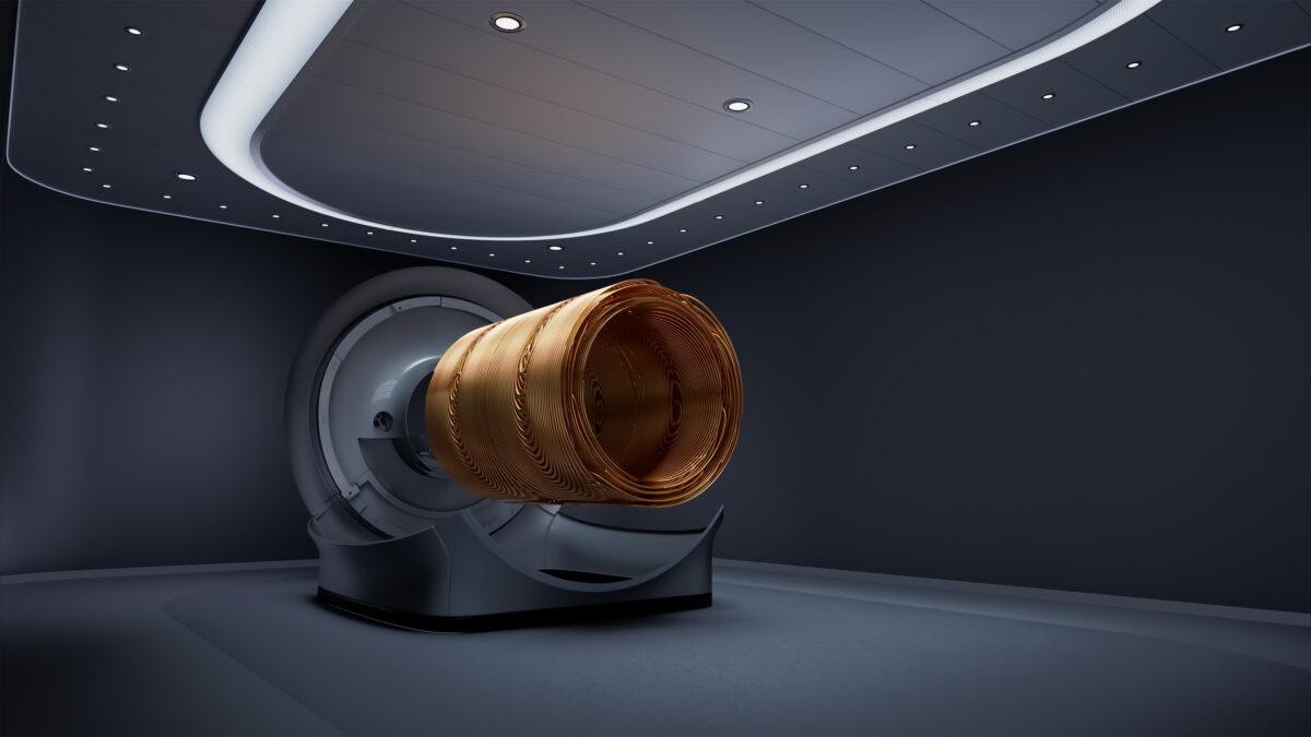
MR 7700 3.0T MR system is the latest break-through MR innovation from Philips and delivers unmatched performance and precision for both research and advanced clinical diagnostics, helping to address the Quadruple Aim.
The XP gradients of MR 7700 3.0T MR system provide high accuracy to support a confident diagnosis for every patient with Philips’ highest quality diffusion imaging [2] and advanced neuroscience, supporting improved patient care and lower costs. MR 7700 expands scanning capabilities with a fully integrated multi-nuclei imaging and spectroscopy solution to explore new clinical pathways, without sacrificing clinical imaging workflow or wide-bore patient comfort, to enhance the experience of both staff and patients.
With the easy to use interface of the MR 7700, scientists and clinicians can now access the scanner without compromising workflow. Driven by seamless integration of multi-nuclei capabilities, the new MR 7700 allows radiologists to image six different clinically relevant nuclei [3] across all anatomies, offering the ability to increase diagnostic confidence and add important metabolic information to MR exams to help tackle complex research programs and improve clinical decision-making to help provide improved patient care. With its AI-driven [1] smart connected imaging, optimized workflows and integrated clinical solutions, MR 7700 helps to improve MR department productivity, enhance patient and staff experience, and deliver high quality diagnostic imaging.
Orchestrating decision-making at pivotal moments in the patient’s care path journey
With all imaging protocols easily accessible via the same intuitive user-friendly interface, MR 7700 contributes to highly efficient radiology workflows and first-time-right scans. Combining high quality imaging with operational efficiency, its XP gradient coils achieve up to 35% higher signal-to-noise ratios and up to 35% shorter scan times [2]. For clinical neurology and neuroscience, radiologists achieve 20% more fMRI volumes [2] and 50% more diffusion tensor imaging (DTI) directions [4], delivering detailed high-resolution images needed to confidently identify and characterize lesions.
“Our highly specialized team of MR physicists are very happy with the ease of use of Philips’ MR 7700 system, especially the implementation of new imaging protocols which is quite simple,” said Prof. Walter Heindel, Professor of Radiology and Chairman of the Department of Radiology at the University Hospital Münster in Germany. “The low effort required for modifying scan parameters and protocols supports fast and easy experimentation with imaging techniques. These latest features clearly help improve our patient and staff experience and distinguish Philips as one of the main reasons we choose this system.”
AI-driven technology helps boost neuroscience excellence
MR 7700 further demonstrates Philips’ commitment to deliver smart diagnostic systems for first-time right diagnosis through clinically relevant and intelligent diagnostics. Clinicians can leverage the MR 7700’s ultra-high gradients and in-built artificial intelligence, such as its AI-driven touchless patient sensing and motion detection, to address the upswing in patient volumes already being experienced by many of today’s radiology departments. Researchers can also leverage the accuracy, power, and endurance of the MR 7700’s XP gradients to help facilitate demanding research programs to boost neuroscience excellence.
“Enhanced with ultra-high gradients and artificial intelligence, Philips MR 7700 is built to deliver on the high-quality clinical performance expectations of today, and to facilitate the most demanding and promising research programs that will help drive the future of MR imaging, without sacrificing workflow efficiency or wide-bore patient comfort,” said Arjen Radder, General Manager of Magnetic Resonance and Diagnostic X-ray at Philips. “Combined with its AI-driven smart connected imaging capabilities, optimized workflows and integrated clinical solutions help meet the quadruple aim of healthcare by improving MR department productivity, enhancing the patient and staff experience, helping to deliver high-quality diagnostic outcomes.”
Connecting and integrating workflows to drive clinical and operational efficiency
MR 7700 leverages the automated technology of SmartWorkflow to guide and coach where required. In addition to conventional proton magnetic resonance imaging and spectroscopy, MR 7700 has in-built MR protocols to combine proton imaging with imaging based on sodium, phosphorus, carbon, fluorine, and xenon [3]. This multi-nuclei capability makes MR 7700 suitable for enhanced anatomical and metabolic/functional imaging in areas such as neurology, pulmonology, and oncology to help deliver fast, and confident definitive decision-making.
Via integration of a dual-tuned head coil, both multi-nuclei and conventional proton MR exams can be performed with the same coil and within the same user interface to help increase workflow efficiencies, while AI-assisted workflow solutions keep exams on schedule to help improve both patient and staff experience. Finally, with access to proactive remote monitoring, remote diagnostics and remote field support, more than 500 parameters of the MR 7700 are monitored from a distance to detect and resolve issues without impacting department operations.
Philips is debuting the MR 7700 at the International Society for Magnetic Resonance in Medicine (ISMRM) annual meeting (May 7-12) in London, and will spotlight the new system at the upcoming European Congress of Radiology (ECR) annual congress in Vienna in July.
This newest MR innovation is part of the Philips Radiology portfolio of scalable high-performance MR systems, including its latest MR 5300 1.5T helium-free in operations MR system, all of which feature intelligent software to automate tasks to help relieve the burden on busy radiology staff and departments. Philips Radiology portfolio delivers patient-centered solutions to reduce anxiety and discomfort; technologist- and radiologist-centered image processing applications for clinical and diagnostic confidence; and enterprise-integrated workflows that streamline the path to precision care.
[1] We embrace the following formal definition of AI from the EU High-Level Expert Group (source: HLEG definition AI)
[2] Compared to Philips’ Ingenia Elition X with Vega HP gradients.
[3] Investigational device for imaging with fluorine (19F) and xenon (129Xe). Limited by federal (or United States) law to investigational use. Clinical imaging with these nuclei requires usage of a cleared drug. No FDA-cleared drugs are currently available for these nuclei.
[4] Compared to Philips DTI/fMRI scans without MultiBand SENSE.


