Radboud University Medical Center Study Sets New Milestone with 90% Diagnostic Accuracy of Lung Nodules Suspected of Cancer Using Philips Technology for Bronchoscopic Procesures

Hospital achieves the world-first level of improvement in the diagnostic accuracy of navigation bronchoscopy using Philips Lung Suite combined with the company’s Image-Guided Therapy System – Azurion

Royal Philips (NYSE: PHG, AEX: PHIA), a global leader in health technology, and Radboud university medical center (Radboudumc), a leading Dutch academic research and teaching hospital (Nijmegen, the Netherlands), have announced the positive results of a clinical study conducted by Radboudumc aimed at setting a new standard of safety and accuracy in the diagnosis of small peripheral lung lesions. The study outcomes represent a promising milestone in the global innovation effort to set a new clinical standard for detecting lung cancer at an early stage, potentially leading to improved prognoses for patients.

Radboudumc’s observational study reported the diagnostic accuracy and procedural radiation dose for patients undergoing an endobronchial lung biopsy supported by Philips Lung Suite – a solution that uses 3D imaging with augmented fluoroscopy to support high precision diagnosis and minimally-invasive therapy in one room. Diagnostic accuracy of 90% was reported while reducing the average total effective radiation dose per procedure by more than half from 47.5 Gy·cm2 (effective dose: 14.3 mSv) to 25.4 Gy·cm2 (effective dose: 5.8 mSv). The median long-axis diameter of the 248 lesions navigated to during the study was 13 mm (range 5-65mm). The results of the study were published in the October 2021 issue of the Journal of Bronchology & Interventional Pulmonology: Volume 28 – Issue 4 [1].

“The airways and lungs are very challenging places to biopsy, so we need good navigation to make sure we reach the target location,” said Dr. Erik van der Heijden, Pulmonologist and Associate Professor of Interventional Pulmonary Diseases at Radboudumc, who led the study. “Philips’ 3D imaging solution allows us to create real-time 3D visualizations enabling us to follow the correct path from different angles to where we need to be, which is particularly useful for very small abnormalities. Our study confirmed that it increases the accuracy and safety of the biopsy procedure, improving results for patients. Philips’ technology also offers the promising outlook that we could not only diagnose but also immediately treat these early-stage patients using novel procedures such as tumor ablation.”

Lung cancer – the leading cause of cancer death

Lung cancer kills around 1.7 million people a year worldwide. That makes it the leading cause of cancer death globally [2], accounting for greater loss of life than breast, colon, and prostate cancer combined. While early diagnosis and treatment are critical to better outcomes and quality of life, the majority of lung cancers are currently diagnosed at a late stage, with minimal chance of a surgical cure.

Early detection and diagnosis key to survivability

The research being carried out at Radboudumc focuses on the diagnosis of early-stage lung cancer lesions, typically detected during routine screening of high-risk patient groups or discovered and reported as so-called ‘incidental findings’ when a patient has a chest scan for some other reason. A tissue sample typically then needs to be obtained that a pathologist can use to make a definitive diagnosis.

Challenging for clinicians to accurately navigate

A new method is image-guided endobronchial lung biopsy, during which a clinician uses live image guidance to maneuver through the airways in the lungs to advance a catheter towards the lesion. However, the airways in the lungs are a complex network that requires accurate navigation to remove the required tissue sample. Until now, image-guided endobronchial lung biopsy has only really been effective when an abnormality or tumor is located in or near one of the larger airways in the lungs, and even then it is challenging for clinicians to accurately navigate when looking at a 2D greyscale fluoroscopy image while the lungs are moving as the patient breathes.

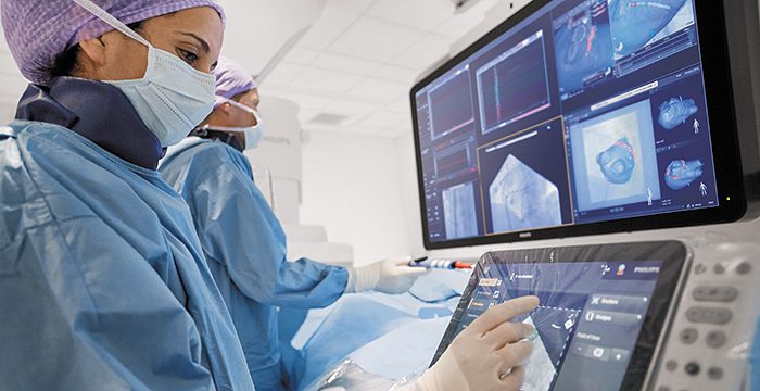
Philips Lung Suite enables all-in-one lung cancer diagnosis and treatment. It provides advanced real-time 3D imaging with augmented fluoroscopy on the company’s Image Guided Therapy System – Azurion, combined with dedicated software. With Philips’ Cone Beam CT imaging, the X-ray detector rotates around the patient to generate a CT-like image in around five seconds, providing clinicians with a high-resolution 3D view of the target lesion and other anatomical structures. This allows the clinician performing the biopsy procedure to be continually guided by high-quality real-time imaging to advance a catheter towards the lesion through a bronchoscope. Once done, its position can be confirmed in real-time using the same imaging modality and a biopsy sample removed.

Philips has a comprehensive portfolio of lung cancer diagnosis and treatment solutions. In addition to Philips Lung Suite, the company’s Lung Cancer Orchestrator provides an integrated lung cancer patient management system for both CT lung screening programs and incidental pulmonary findings programs that manages and monitors patients every step of the way in their lung cancer screening and treatment decision journey.


References

[1] Journal of Bronchology & Interventional Pulmonology: October 2021 – Volume 28 – Issue 4 – p 262-271 doi: 10.1097/LBR.0000000000000783
[2] CA CANCER J CLIN 2021;71:7–33, Cancer Statistics 2021: https://acsjournals.onlinelibrary.wiley.com/doi/epdf/10.3322/caac.21654
Hot this week

Cartessa Aesthetics Partners with Classys to Bring EVERESSE to the U.S. Market

Classys, which is listed on the KOSDAQ, is one of South Korea's most distinguished aesthetic technology manufacturers, with devices distributed in 80+ markets globally. This partnership marks Classys's official entry into the American marketplace, with Cartessa Aesthetics as the exclusive distributor for EVERESSE, launched under the Volnewmer brand in current global markets.

Stryker Launches Next-Generation of SurgiCount+

Now integrated with Stryker's Triton technology, SurgiCount+ addresses two key challenges: retained surgical sponges and blood loss assessment. Integrating these previously separate digital solutions provides the added benefit of a more efficient, streamlined workflow for hospitals notes Stryker.

Nevro Receives CE Mark In Europe for It’s HFX iQ™ Spinal Cord Stimulation System

Nevro notes HFX iQ is the first and only SCS system with artificial intelligence (AI) technology that combines high-frequency (10 kHz) therapy built on landmark evidence that uses ongoing cloud data insights to deliver personalized pain relief

Recor Medical Reports: CMS Grants Distinct TPT Device Code and Category to Recor Medical for Ultrasound Renal Denervation

The approval of TPT offers incremental reimbursement payments for outpatient procedures performed with ultrasound renal denervation for Medicare fee-for-service beneficiaries. It becomes effective January 1, 2025, and is expected to remain effective for up to three years notes Recor Medical.

Jupiter Endovascular Reports | 1st U.S. Patient Treated with Jupiter Shape-shifting Thrombectomy Device

“Navigation challenges during endovascular procedures are often underappreciated and have led to under-adoption of life-saving procedures, such as pulmonary embolectomy. We have purpose-built our Endoportal Control technology to solve these issues and make important endovascular procedures accessible to more clinicians and their patients who can benefit from them,” said Carl J. St. Bernard, Jupiter Endovascular CEO. “This first case in the U.S. could not have gone better, and appears to validate the safety and performance we are seeing in our currently-enrolling European SPIRARE I study.”
Exit mobile version